Scientists in S Korea have cloned 30 human embryos. They hope to obtain cells that could eventually be used to treat disease.
The took the genetic material from normal cells in women donors and combined it with their eggs.
This was done at the Seoul National University by Suk Hwanf and his team.
The embryos were then developed to produce stem cells. These stem cells can divide into any tissue in the body.
These cells will (it is hoped) replace those which have failed, for example, in patients with Alzheimer's disease.
'Because these cells carry the nuclear genome of the individual, after differentiation they could be expected to be transplanted without immune rejection for treatment of degenerative disorders. Our approach opens the door for the use of these specially developed cells in transplantation medicine.' said Professor Hwang.
His research is being published online by the journal Science (Science Express web site).
Mice proof
There have been claims in the past for the creation of human embryo clones to study so-called stem cells - many of them disputed.
But no group has reported producing so many early-stage clones and seen their development progress to such an advanced stage.
The work has also been subjected to the rigorous scrutiny of independent scientists before publication in a major journal.
'These are the most advanced human embryo clones yet produced,' Professor Hwang told BBC News Online.
The team says it sought approval for its work from an ethical review board and obtained informed consent from its women donors before proceeding with the work.
Thirty embryos
The team tells Science Express how it used 242 eggs in its experiments taken from 16 women.
In each case, material was transferred from the nucleus of a non-reproductive (somatic) cell, containing the woman's genetic blueprint, into a nucleus-free egg from the same donor.
Following this transfer, factors within the host egg's exterior, or cytoplasm, are believed to have reprogrammed the new nuclear contents by activating versatile embryonic genes, while silencing the more limited adult somatic cell genes.
In total, 30 embryos - exact genetic copies of their female donors - were then cultured to the so-called blastocyst stage at which stem cells could be extracted.
These special cells were seen to divide into all three of the main tissue types found in the human body, the researchers report.
The cells were even transplanted into mice to show they could differentiate into still more specific cell types, offering further proof of their 'pluripotency'.
The stated intention is to study human embryonic stem cells to see how they could be used as a therapeutic tool to treat disorders, such as diabetes, osteoarthritis, and Parkinson's disease, among others, in which tissues in the body have begun to fail.
Non-egg future
Editor-in-chief of the journal Science, Donald Kennedy, said: 'The potential for embryonic stem cells is enormous, but researchers still must overcome significant scientific hurdles.'
And he added: 'These results seem promising. But, it's important to remember that cell and tissue transplantation and gene therapy are still emerging technologies, and it may be years yet before embryonic stem cells can be used in transplantation medicine.'
Addressing ethical concerns, he also called for a worldwide ban on activities which would seek to use this technology to create living children.
Professor Hwang, whose expertise has been developed in animal cloning, said any attempt to produce a baby would be 'crazy'.
'We will never try to produce cloned human beings,' he said.
'During animal cloning, we experienced so many difficulties and dangers with deformities, especially in the internal organs.'
Commenting on the Korean work, Roger Pedersen, professor of regenerative medicine, at the University of Cambridge, UK, told BBC News Online: 'The present work has substantially advanced the cause of generating transplantable tissues that exactly match the patient's own immune system.
'These researchers' findings also make it possible to learn how to reprogramme the human genome to an embryonic state.
'This will likely accelerate the development of alternative ways of reprogramming human cells, which could in the future diminish the need to use human eggs for this purpose.'

American Association for the Advancement of Science
The embargo on this press release has been lifted ahead of schedule.
SEATTLE, WA--New research - published by Science Magazine within the Science Express Web site and released today at the 2004 AAAS Annual Meeting -- may be a first step toward methods for treating diabetes, osteoarthritis, Parkinson's and other diseases, by producing replacement cells unlikely to trigger immune-system rejection.
Transplantation medicine based on stem cells remains a distant hope for now, Science editors cautioned. But, the Science study describes intriguing early results:
For the first time, researchers have reported the development of versatile 'pluripotent' human embryonic stem cells, potentially capable of becoming any cell in the body, from a cloned human blastocyst.
The stem cells were harvested from a blastocyst produced by transferring the nucleus of a non-reproductive ('somatic') cell, containing a woman's genetic blueprint, into a nucleus-free egg from the same donor.
Following this transfer, factors within the host egg's exterior, or cytoplasm, reprogrammed its new nuclear contents by activating versatile embryonic genes, while silencing the more limited adult somatic cell genes. Researchers were then able to collect embryonic stem cells from the resulting cell mass inside the cloned blastocysts.
In theory: 'Because these cells carry the nuclear genome of the individual, after differentiation they could be expected to be transplanted without immune rejection for treatment of degenerative disorders,' reported Woo Suk Hwang of Seoul National University in Korea.
'Our approach opens the door for the use of these specially developed cells in transplantation medicine.'
Embryonic stem cells have previously been produced with cells from mice using the same method, called 'somatic cell nuclear transfer.' But, achieving this trick with human cells posed unique challenges, said Donald Kennedy, Science's Editor-in-Chief.
The researchers attribute their apparent success to the use of extremely fresh donor eggs, stringent timing protocols, and a special method for gently extruding rather than suctioning the DNA-spindle complex from eggs. Suctioning the DNA may damage spindles, possibly causing chromosomal defects called aneuploidy, they noted.
Hwang and colleagues developed the stem cell line, SCNT-hES-1, after collecting 242 eggs from 16 unpaid volunteers who had signed informed-consent agreements. From these eggs, scientists then cultured 30 blastocysts to obtain 20 suitable inner cell masses. By tweaking the amount of time that elapsed between the transfer of the nucleus and the activation of the newly transplanted genetic material, the team was able to optimize their results:
A two-hour delay seemed to work best, so that 20 percent of all reconstructed eggs formed blastocysts. From the inner cell mass of these blastocysts, a single human embryonic stem cell line was obtained.
The resulting stem cells differentiated into all three of the main tissue types that appear at the beginning stages of development, researchers reported. When transplanted into mice, the stem cells differentiated into still more specific cell types, offering further proof of pluripotency.
Interestingly, the research team harvested eggs as well as somatic cells from the same donors: Nuclear material from the somatic cell was transferred into the nucleus-free or enucleated egg of the same woman.
This unusual experimental design may be more effective than person-to-person transfers because it offered greater compatibility between the genetic components that were fused together.
But, were the stem cells truly derived from the transferred nucleus, or were they the result of an accidental 'parthenote'--an artificially induced blastocyst resulting from an egg that began to spontaneously divide?
To support their claim that the resulting stem cells came from the transplanted nucleus, Hwang's team completed DNA fingerprinting analysis, and also checked the expression of imprinted genes.
The results were consistent with stem cells resulting from transplantation.
Many questions remain, Science's Donald Kennedy said: 'The potential for embryonic stem cells is enormous, but researchers still must overcome significant scientific hurdles,' Kennedy remarked.
'These results seem promising. But, it's important to remember that cell and tissue transplantation and gene therapy are still emerging technologies, and it may be years yet before embryonic stem cells can be used in transplantation medicine.'
The research also raises policy and ethical questions, Kennedy noted, since blastocyst-derived stem cells for tissue repair or transplantation might exacerbate pressures on egg donors in some regions.
The prospect of using cloned human blastocysts to produce new embryonic stem cells lines also is likely to provoke further controversy, he added. 'There is widespread consensus among all responsible, mainstream scientists--including the authors of this paper and AAAS, publisher of Science Magazine--that any attempt to clone a human being would be highly dangerous and wrong, and therefore, all reproductive cloning should be banned,' Kennedy said.
'But, the generation of stem cells by somatic cell nuclear transfer methods involving the same individuals may hold promise for advances in transplantation technology that could help people affected by many devastating conditions.'
In addition to Hwang, authors on this Science paper were Young June Ryu, Eul Soon Park, Eu Gene Lee, Hyun Yong Chun, Byeong Chun Lee, Sung Keun Kang, Curie Ahn and Shin Yong Moon, all of Seoul National University; as well as Jong Hyuk Park and Sun Jong Kim of Mizmedi Hospital in Seoul; Ja Min Koo of Gachon Medical School; Jung Hye Hwang of Hanyang University; Ky Young Park of Sunchon National University; and Jose B. Cibelli of Michigan State University.
The American Association for the Advancement of Science (AAAS) is the world's largest general scientific society, and publisher of the journal, Science (www.sciencemag.org). AAAS was founded in 1848, and reports some 265 affiliated societies and academies of science, serving 10 million individuals. Science has the largest paid circulation of any peer-reviewed general science journal in the world, with an estimated total readership of one million. The non-profit AAAS (www.aaas.org) is open to all and fulfills its mission to 'advance science and serve society' through initiatives in science policy; international programs; science education; and more. For the latest research news, log onto EurekAlert!, www.eurekalert.org, the premier science-news Web site, a service of AAAS.
MEDIA NOTE: A newsbriefing on this research will take place at 11:00 a.m. Pacific Time, Thursday, 12 February, during the AAAS Annual Meeting in Seattle, in the Eliza Amphitheater, Grand Hyatt. Further, these and other speakers will take part in a symposium titled, 'Stem Cell Science in the Service of Society,' at 2:30 p.m. Monday, 16 February, Sheraton Hotel, Second Floor, Grand Ballroom C. Press registration is in the AAAS Press Center in Leonesa I of the Grand Hyatt Hotel.
AAAS is the world's largest general scientific society, dedicated to 'Advancing science � Serving society.'
Contact: Ginger Pinholster
gpinhols@aaas.org
206-774-6330

Stem cell research has been given a boost after a new study shows a link between the controversial technique and couples undergoing IVF treatment.
The survey found that 57% of IVF couples would consent to their surplus embryos being used for stem cell research - because they receive better information on the issue.
Medical experts say the number of couples handing over surplus embryos depends upon the quality of information about the needs and benefits of stem cell research given.
Stem cell research has been the target of huge criticism from pro-life campaigners and religious communities who believe embryos are already a human life and should be treated as such.
Professor Alison Murdoch, chairman of the British Fertility Society said: 'Our results are encouraging as they show that couples undergoing IVF understand the need and benefits of embryo research, probably because they have access to good information that the majority of the population do not.
'When people understand this issue they tend to look on it favourably.
'Scientists should not be afraid of engaging the public on this issue.'
Clare Brown, chief executive of Infertility Network UK, added: 'The results of this study highlight the fact that couples are keen to assist others while going through what is an extremely difficult, both physically and emotionally, treatment for themselves personally.
'The key word for couples is obviously `information'.'
Meanwhile, new research shows that sperm counts have dropped by almost a third in a decade.
Drug use, alcohol, smoking and obesity are among the factors most frequently blamed.
Each attempt at IVF treatment costs about �3,000 and the NHS funds only one in five of these attempts.
It is expected that a new recommendation will entitle thousands of women aged between 23 and 39 to free IVF treatment.

A group of researchers from The Scripps Research Institute (USA) has identified a small synthetic molecule that can induce a cell to undergo dedifferentiation--to move backwards developmentally from its current state to form its own precursor cell.
This compound, named reversine, causes cells which are normally programmed to form muscles to undergo reverse differentiation--retreat along their differentiation pathway and turn into precursor cells.
These precursor cells are multipotent; that is, they have the potential to become different cell types. Thus, reversine represents a potentially useful tool for generating unlimited supply of such precursors, which subsequently can be converted to other cell types, such as bone or cartilage.
'This [type of approach] has the potential to make stem cell research more practical,' says Sheng Ding, Ph.D. 'This will allow you to derive stem-like cells from your own mature cells, avoiding the technical and ethical issues associated with embryonic stem cells.'
Ding, who is an assistant professor in the chemistry department at Scripps Research conducted the study--to be published in an upcoming issue of the Journal of the American Chemical Society--with Peter G. Schultz, Ph.D., who is a professor of chemistry and Scripps Family Chair of Scripps Research's Skaggs Institute of Chemical Biology, and their colleagues.
Regenerative Medicine and Stem Cell Therapy
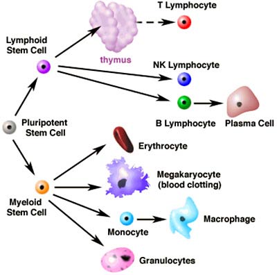
Stem cells have huge potential in medicine because they have the ability to differentiate into many different cell types--potentially providing doctors with the ability to produce cells that have been permanently lost by a patient.
For instance, the damage of neurodegenerative diseases like Parkinson's, in which dopaminergic neurons in the brain are lost, may be ameliorated by regenerating neurons.
Another example of a potential medical application is Type 1 diabetes, an autoimmune condition in which pancreatic islet cells are destroyed by the body's immune system. Because stem cells have the power to differentiate into islet cells, stem cell therapy could potentially cure this chronic condition.
However bright this promise, many barriers must be overcome before stem cells can be used in medicine. Stem cell therapy would be most effective if you could use your own stem cells, since using one's own cells would avoid potential complications from immune rejection of foreign cells.
However, in general it has proven very difficult to isolate and propagate stem cells from adults. Embryonic stem cells (ESCs) offer an alternative, but face both practical and ethical hurdles associated with the source of cells as well as methods for controlling the differentiation of ESCs.
A third approach is to use one's own specialized cells and dedifferentiate them.
Normally, cells develop along a pathway of increasing specialization. Muscles, for instance, develop after embryonic stem cells develop into 'mesenchymal' progenitor cells, which then develop into 'myogenic' cells. These muscle cells fuse and form the fibrous bundles we know as muscles.
In humans and other mammals, these developmental events are irreversible, and in this sense, cell development resembles a family tree. One wouldn't expect a muscle cell to develop into a progenitor cell any more than one would expect a woman to give birth to her own mother.
However, such phenomena do happen in nature from time to time.
Some amphibians have the ability to regenerate body parts that are severed by using dedifferentiation.
When the unlucky amphibian loses a limb or its tail, the cells at the site of the wound will undergo dedifferentiation and form progenitor cells, which will then multiply and redifferentiate into specialized cells as they form an identical replacement to the missing limb or tail.
In humans, the liver is unique in its regenerative capacity, possibly also involving dedifferentiation mechanism.
The Scripps Research scientists hope to find ways of mimicking this natural regeneration by finding chemicals that will allow them to develop efficient dedifferentiation processes whereby healthy, abundant, and easily accessible adult cells could be used to generate stem-like precursor cells, from which they could make different types of functional cells for repair of damaged tissues.
Reversine is one of the first steps in this process.
However, tissue regeneration is years away at best, and at the moment, Schultz and Ding are still working on understanding the exact biochemical mechanism whereby reversine causes the muscle cells to dedifferentiate into their progenitors, as well as attempting to improve the efficiency of the process.
'This [type of research] may ultimately facilitate development of small molecule therapeutics for stimulating the body's own regeneration,' says Ding. 'They are the future regenerative medicine.'
The article, 'Dedifferentiation of Lineage-Committed Cells by a Small Molecule' is authored by Shuibing Chen, Qisheng Zhang, Xu Wu, Peter G. Schultz, and Sheng Ding and is available to online subscribers of the Journal of the American Chemical Society at: http://pubs.acs.org/cgi-bin/asap.cgi/jacsat/asap/html/ja037390k.html. The article will also be published in an upcoming issue of JACS.
This work was supported by The Skaggs Institute for Research and the Novartis Research Foundation.

Studies in zebrafish lead to better understanding of blood formation and leukemia development
Boston--Researchers at Children's Hospital Boston have isolated a gene responsible for making blood stem cells. The findings appear in today's issue of the journal Nature. The gene, called cdx4, is responsible for establishing the location of blood cell formation in the developing embryo.
Cdx4 works by altering the expression of HOX genes, which are involved in making the body plan. Surprisingly, the authors found that overexpression of cdx4 in zebrafish embryos, or in mouse embryonic stem cells, induces the new production of early blood cells.
'We have been searching for genes in the zebrafish that participate in making blood stem cells,' according to lead author, Leonard Zon, MD., of Children's Hospital Boston.
'Now that we have these genes, we are one step closer to growing more blood stem cells. This will be potentially useful for patients with severe congenital anemias or bone marrow transplantation for cancer,' adds Zon.
Scientists studied a mutant that had a severe anemia because it had few blood stem cells, and also had a tail defect. The zebrafish mutants generally die within seven to ten days after fertilization.
They discovered the mutation in the cdx4 gene, which is associated with the early blood deficiency as well as abnormal developmental patterning, including aberrant hox gene expression.
When researchers injected the mutants with hox genes, such as hoxb7a and hoxa9a, it resulted in almost complete rescue of the deficient blood cells. Another hox gene, hoxb6b showed some improvement, but hoxb8a did not have any effect on the blood defect.
Researchers believe this shows blood cell development is dependent on the proper expression of these hox genes, and that overexpression of these genes can reverse a fatal deficiency in these blood cells.
'These zebrafish findings will allow us to better understand normal blood development, with the hopes of eventually developing more effective treatments for these devastating blood disorders such as leukemia,' says Zon.
Children's Hospital Boston is home to the world's largest research enterprise based at a pediatric medical center, where its discoveries have benefited both children and adults for over 100 years. More than 500 scientists, including seven members of the National Academy of Sciences, nine members of the Institute of Medicine and nine members of the Howard Hughes Medical Institute comprise Children's research community. Founded in 1869 as a 20-bed hospital for children, Children's Hospital Boston today is a 300-bed comprehensive center for pediatric and adolescent health care grounded in the values of excellence in patient care and sensitivity to the complex needs and diversity of children and families. It is also the primary pediatric teaching affiliate of Harvard Medical School. For more information about the hospital visit: www.childrenshospital.org.
Embryonic stem cells have been encouraged to grow into sperm cells for the first time, Japanese scientists report.
The work is very preliminary, and was done in the laboratory with mouse stem cells. The next step would be to see if it can be repeated in live animals.
Stem cells are the basic building blocks of animals, forming in the new embryo and later developing into the various organs and tissues as the fetus grows.
Researchers have grown stem cells into many other types of cells, including egg cells, but this is the first time a sperm cell has been developed, the scientists said.
The work was headed by Toshiaki Noce of Mitsubishi Kagaku Institute of Life Science in Japan. The results are reported in this week's online issue of Proceedings of the National Academy of Science.
Noce and his team incubated the stem cells with other cells that produce a protein called BMP4, which is known to stimulate formation of sperm cells during the development of an embryo. In their laboratory, some of the stem cells began developing into sperm cells within one day, a process that takes three days in the embryo.
Bert Vogelstein, a professor at Johns Hopkins University who headed an Academy panel on stem cells, said the work is 'novel and provides a wonderful example of how new technologies can provide diverse cell types in the test tube that may prove useful for biomedical applications in the future.'
Growing stem cells into other tissues has been hailed as a source of major potential therapies in the future. However, the process is controversial because many stem cells are harvested from discarded embryos.
The Bush administration has limited federal funding for stem cell research to lines of cells that already exist, although new stem cell lines can be developed using private funds and in some other countries.
Study adds to evidence of adult stem cells' promising therapeutic role
BURLINGTON, VT (USA) - For the first time, researchers have demonstrated that adult human stem cell transplantation results in spontaneous cell regeneration in damaged lung tissue.
Published in the August 1 issue of the American Journal of Respiratory and Critical Care Medicine, the study further supports an existing body of research that suggests blood- and marrow-derived stem cells have the capacity to become many different human tissues.
'Many of the body's tissues once thought to be only locally regenerative may, in fact, be actively replaced by circulating stem cells after hematopoietic or blood-forming stem cell transplantation,' says lead author Benjamin Suratt, M.D., assistant professor of medicine and Vermont Lung Center researcher at the University of Vermont College of Medicine.
'This finding is of note not only for its novelty as a regenerative mechanism of the lung, but also for its vast therapeutic implications for any number of lung diseases.'
According to Suratt, the study's findings indicate that circulating stem cells are going into organ tissue and repairing damage, which could have a huge impact on the treatment of such devastating lung diseases as emphysema or cystic fibrosis.
Supported by funding from the National Institutes of Health and a National Center for Research Resources Centers for Biomedical Research Excellence grant, Suratt and his colleagues are currently looking further into what types of cells have the capacity to differentiate and generate a different type of cell, and whether these cells might be used to treat cystic fibrosis.
For more information on research taking place at the Vermont Lung Center at the University of Vermont, go to www.vermontlung.org
To link to the article abstract, go to:
ajrccm.atsjournals.org/cgi/content/abstract/168/3/318

บทความที่ใหม่กว่า บทความที่เก่ากว่า หน้าแรก
